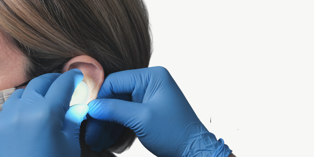
The importance of using proper illumination and magnification while performing cerumen management cannot be stressed enough. It all starts with conducting a thorough cerumen impaction assessment using an otoscope or video otoscope to determine the type of cerumen present, and any modifying factors or contraindications to proceeding with treatment.
Following the initial examination, if the decision is to proceed, further illumination or magnification may be required depending on the method of removal chosen to extract the impaction. Irrigation or the use of a tool like the EarWay Pro requires minimal illumination during the actual procedure. During the procedure, the device used for the initial assessment can be used to monitor progress or the success of the extraction at completion.
If, however, the method selected uses tools such as curettes or micro-suction, then proper illumination and sometimes magnification, are required. Good lighting and magnification are needed both for patient safety and for accurate handling of the tools in the external ear canal. Illumination and magnification systems that allow hands-free operations during cerumen removal are preferred. In a perfect world, anyone performing cerumen management would have access to a microscope, but these are cost-prohibitive for most clinics and are not portable, so our focus here is on portable, head-worn systems.
Headlamps
If your preference or requirement is for illumination only, there are several choices for your consideration. A simple forehead reflection mirror can provide additional illumination down the ear canal. Forehead reflection mirrors are inexpensive, low tech and very portable, requiring only some type of light source in the room to reflect off the mirror. A head-worn light or headlamp, like the ones people use for camping, can also offer a low-tech solution and the freedom to move around while maintaining the focus of the beam of light in the direction you are looking.

Head Loupes
My preference is to use a product that can provide both good illumination and magnification during manual or micro-suction extraction procedures of cerumen. There are several medical-grade products on the market that are used in dentistry, ENT, or by other surgical professionals. These products have been designed to provide excellent lighting and magnification required for delicate procedures. Companies such as Heine and Zeiss are well known for their operating head-worn systems, also called head loupes. As these are medical-grade systems, they tend to have higher costs than a similar system designed for a jeweller or hobbyist.
Head loupes can provide excellent levels of magnification (up to 6X) and lighting. Costs have quite a range, but one of the major drawbacks of most head loupe systems is that they make it difficult, if not impossible, to achieve a binocular focal point down the ear canal. Clinicians, myself included, find that when using a head loupe, reliance on monocular vision is not ideal to see down the ear canal. This is because depth perception is lost when it is needed the most for the accurate positioning and manipulation of implements in the ear canal.
The Vorotek O Scope
A type of head-worn illumination and magnification system that I have become knowledgeable of, called the Vorotek O Scope, from Australia, was designed by an ENT surgeon to achieve a binocular focal point down a tube, i.e., the ear canal. This head-worn system (Vorotek calls it a head-worn microscope) allows for hands-free operation, excellent illumination, and a level of magnification adequate for cerumen extraction purposes. The one drawback of this system is limited magnification levels (roughly 1.5X) compared to head loupes that can offer more. The cost of the O-Scope is less than premium head loupes, making it an excellent choice for audiology clinics.


