Hearing healthcare providers will frequently encounter conditions of the external ear that require special consideration when conducting hearing evaluations, ear impressions, hearing aid fittings, and cerumen management.
Here we discuss the most common conditions that are encountered by hearing care professionals during otoscopy. We will also discuss the appropriate course of action to address the various conditions outlined. It should be noted, however, that unless it is within your scope of practice, you will not be making a diagnosis of a specific condition. It is nonetheless important to have some familiarity with the presenting condition so that appropriate action can be taken. With that in mind, let’s begin.
Common Ear Conditions
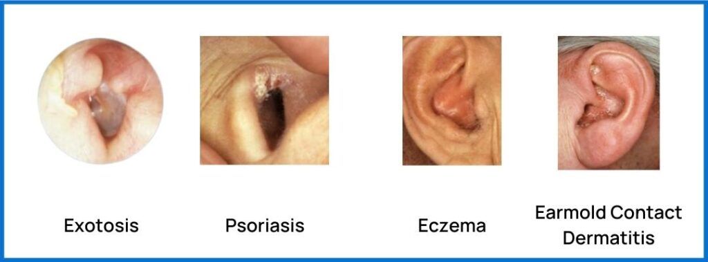
Exostosis
What It Is: The formation of new bone on the surface of a bone, is generally caused by exposure to cold water and cold wind.
What To Look For: Multiple smooth round protrusions, white in colour, will be seen during otoscopic examination.
Prevalence: 0.63% in the general population, 73.5% in surfers.
Cerumen Management Indications: Can create additional sensitivity for your patient during cerumen removal. If using instrumentation do not touch the growths; for irrigation using distilled water is important, and irrigation should not be performed on anything other than mild exostosis.
More Info
Psoriasis
What It Is: An autoimmune disease that causes the skin’s life cycle to accelerate.
What To Look For: Small to large areas of irritated skin that won’t heal, dry or cracked skin that bleeds, and temporary hearing loss from blocked ears.
Prevalence: 0.6% in the general population.
Cerumen Management Indications: Only proceed with cerumen management following medical clearance.
More Info
Dermatitis
What It Is: A general term that describes a skin irritation or inflammation, which can be infectious or non-infectious. There are two types of dermatitis that are frequently seen by hearing care professionals:
- Eczema
- What To Look For: Dryness, redness, scaliness, itchiness and cracked skin on or behind the ears. Discharge is also common. Symptoms are usually worse in the winter.
- Prevalence: About 1 in 10 individuals over a lifetime (general eczema, not ear-specific).
- Cerumen Management Indications: Cerumen management should not be performed on ears with eczema without medical clearance as they are more prone to infection.
- More Info
- Earmold Contact Dermatitis
- What To Look For: Red, itchy rash, swelling.
- Prevalence: Unknown
- Cerumen Management Indications: The type of earmold material should be changed to a hypoallergenic material. If that clears it up then you are okay to proceed with cerumen removal, if not then a medical referral for management of contact dermatitis is warranted.
- More Info
Common Infections of the Ear
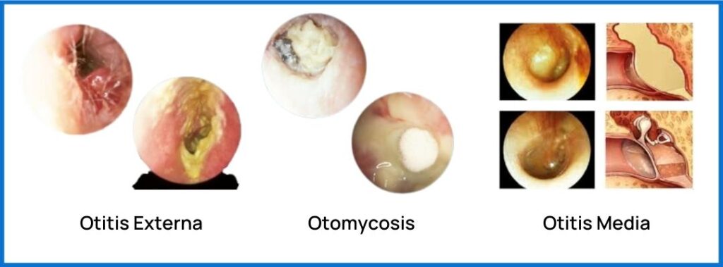
Otitis Externa
What It Is: Inflammation of the ear canal skin, typically bacterial in nature but can also be fungal; there are three sub-types:
- Acute: Refers to a single episode, typically lasting a short period of time following successful medical treatment
- Chronic: Refers to ongoing episodes, typically lasting more than 3 months.
- Necrotizing (aka Malignant): A complication of otitis externa; an invasive infection of the external auditory canal and skull base. It is potentially life-threatening and occurs primarily in older persons who have diabetes mellitus or another immunocompromising condition.
What To Look For: Ear canal inflammation, itching, discharge and pain.
Prevalence: Prevalence is 10% of the population over their lifetime, affecting adults more commonly than children.
Cerumen Management Indications: Cerumen management is contraindicated, and medical referral is warranted.
More Info
Otomycosis
What It Is: Fungal infection of the external ear, middle ear, or mastoid cavity.
What To Look For: Symptoms include pain, fullness, discharge, and itchiness. Look for yellow bumpy dots.
Prevalence: About 10% of all cases of otitis externa are fungal.
Cerumen Management Indications: Cerumen management is contraindicated; medical referral is warranted.
More Info
Otitis media
What It Is: A group of inflammatory diseases of the middle ear that is often a complication of eustachian tube dysfunction where the middle ear is not being ventilated and cleared adequately. Can be infections or non-infectious (also called OM with effusion). There are 2 subtypes:
- Acute: An infection of rapid onset that usually presents with ear pain.
- Chronic: Refers to ongoing episodes, lasting more than 3 months.
What To Look For: inflammation, fullness, pain, potential fever, and conductive hearing loss, typically in the low frequencies. Flat tympanogram.
Prevalence: About 80% of children will suffer at least one episode of acute otitis media; more common in children than adults as the eustachian tubes are not fully matured until age 7 or 8 as they lie more horizontally.
Cerumen Management Indications: Otitis media is a contraindication to cerumen removal and a red flag for medical referral when observed.
More Info
Common Tympanic Membrane Pathologies
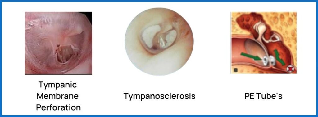
Perforation
What It Is: A hole or tear in the tympanic membrane (TM) tissue separating the middle ear from the external ear. This can be due to a puncture wound or bursting of the tissues because of an infection. Pressure Equalization (or PE) Tubes are tiny hollow tubes that are surgically placed in the tympanic membrane to assist with ventilation of the middle ear space for the treatment of chronic otitis media.
What To Look For: Visible hole in TM, history of otitis media, pain, conductive hearing loss, flat tympanogram with high ear canal volume. For PE tubes, they should be visible unless obstructed by cerumen.
Prevalence: incidence of traumatic TM perforation is reported in the literature as affecting 0.68% of the population.
Cerumen Management Indications: Contraindicates certain cerumen removal procedures, such as the use of softening agents and irrigation. For some licences, cerumen removal is not permitted when perforations of any type are present.
More Info
Tympanosclerosis
What It Is: A condition caused by hyalinization and subsequent calcification of subepithelial connective tissue of the tympanic membrane. Healed perforations can also cause tympanosclerosis.
What To Look For: the visual appearance of white spots or clouds floating on the tympanic membrane that can result in a conductive hearing loss.
Prevalence: 19% amongst the population with chronic suppurative otitis media.
Cerumen Management Indications: The use of irrigation is not advisable due to the reduced capacity for the tympanic membrane to take tensile pressure.
More Info
Common Ear Traumas & Foreign Bodies
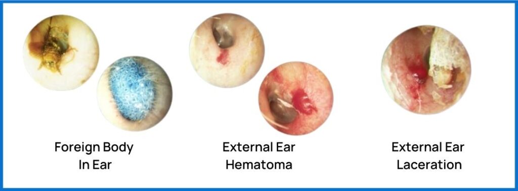
Foreign Bodies & Objects in the Ear Canal
What It Is: A foreign object that has somehow made its way into the ear canal.
What To Look For: Hearing aid domes are often common, but clear and hard to visualize. Insects can integrate with cerumen and be hard to visualize, high magnification video otoscopes can be helpful. Earplugs are often brightly coloured and are easier to visualize.
Prevalence: About 4%; predominantly in children.
Cerumen Management Indications: Alligator forceps or suction would be best tools to consider in these cases. Removing objects from a child’s ear canal may not be in your scope of practice. In this case, a medical referral is warranted.
More Info
Trauma of the External Ear
What It Is: Injury to the tissues lining the canal. There are two types which commonly affect the ears:
- External Ear Hematoma – When a blood vessel leaks into the surrounding tissue.
- External Ear Laceration – A wound that is produced by the tearing of soft body tissue.
What To Look For: A hematoma will present as a blood blister and a laceration will look like a wound with broken skin.
Prevalence: Unknown in general; 2.4% among those who practice ear cleaning using cotton buds.
Cerumen Management Indications: Postponement of cerumen removal is advised until the condition has been resolved.
More Info
Common Cancers & Growths of the External Ear
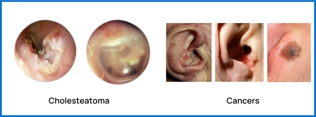
Cholesteatoma
What It Is: An abnormal, noncancerous skin growth in the middle ear behind the eardrum, it may be congenital (present at birth), or caused by repeated middle ear infections.
What To Look For: Discharge from the ear, hearing loss, dizziness, potential facial paralysis, foul odour.
Prevalence: The annual incidence of acquired cholesteatoma ranges from approximately 9 to 12.6 cases per 100,000 adults and from 3 to 15 cases per 100,000 children, about 70-96% of cholesteatomas are acquired.
Cerumen Management Indications: Contraindicated; immediate referral to an otolaryngologist for medical intervention is required.
More Info
Cancer of the External Ear
What It Is: Basal cell skin carcinoma, squamous cell carcinoma, and melanoma.
What To Look For: Oozing and bleeding is a common presentation from a crusted-over wound that does not readily heal. Present as a flat, rough, crusty spot of skin. The lesion will appear to be raised on the surface of the skin and it malignantly evolves anywhere on the outer ear. When touched, the lesion will be firm. Melanomas are mole-like, and in some cases, they develop from moles and may appear red, pink, blue, white, purple, brown or black in colour.
Prevalence: Approximately 300 people in the US are diagnosed with ear cancers each year.
Cerumen Management Indications: Cerumen removal should not be undertaken without medical clearance.
More Info
Rarer Conditions & Infections
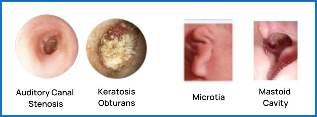
Auditory Canal Stenosis
What It Is: A narrowing of the ear canal; usually congenital or occasionally caused by repeated infections, inflammation, or trauma.
What To Look For: A canal diameter of less than 4mm is the clinically suggested diagnostic criteria for stenosis.
Prevalence: 0.01 to 0.1% for congenital and for acquired, it is even less.
Cerumen Management Indications: We recommend that cerumen management for this condition only be performed by an Otolaryngologist. If it is within your scope of practice as an allied healthcare professional, then it should only be performed with medical clearance using micro-suction. Needs ENT investigation as there is a high risk for cholesteatoma.
More Info
Keratosis Obturans
What It Is: A buildup of keratin, a protein in skin cells, in the ear canal.
What To Look For: Presents like impacted cerumen, however, can cause significant pain upon attempted removal.
Prevalence: Extremely rare, no statistics exist.
Cerumen Management Indications: This condition should be referred for medical investigation and treatment. Do not proceed with cerumen management unless it is within your scope of practice.
More Info
Atresia, Microtia & Anotia
What It Is: Abnormalities or deformities of the external ear are often co-occurring so we will discuss them together.
- Aural Atresia (AA) is a congenital absence or underdevelopment of the external auditory canal.
- Microtia is a congenital deformity where the pinna is underdeveloped or incompletely formed.
- Anotia is the absence of an external ear.
What To Look For: Deformed outer ear. These are congenital and normally diagnosed at birth.
Prevalence: Incidence of microtia is reported to be 0.5 to 3 per 10 000 live births, and AA was reported in 55% to 93% among individuals with microtia.
Cerumen Management Indications: Patients are likely monitored by otolaryngologists and would not be attending your clinic for cerumen management of the impaired ear(s).
More Info
Mastoid Cavity
What It Is: Result of a canal wall down mastoidectomy, a surgery to remove infected cells in the hollow, air-filled spaces in the skull behind the ear within the mastoid bone.
What to Look For: A hollow bowl off the superior-posterior wall of the ear canal; obvious signs of ear surgery; history of mastoiditis.
Prevalence: between 30,000 and 60,000 mastoidectomies are performed each year in the United States as of 2002.
Cerumen Management Indications: You should not be performing cerumen management in these cases. These cases should be, and typically are managed by an otolaryngologist who will regularly see the patient for ongoing cerumen removal and debridement.
More Info
It is important to understand the limitations of your scope of practice, but also to use your clinical judgement and err on the side of caution by referring many of these conditions for medical evaluation, particularly if they have not yet been investigated. If you would like to learn more about cerumen management or become certified in cerumen management check out our online course. Please contact us at info@pacificaudiologygroup.com if you have any questions.
References
- Wong BJF, Cervantes W, Doyle KJ, et al. Prevalence of External Auditory Canal Exostoses in Surfers. Arch Otolaryngol Head Neck Surg. 1999;125(9):969–972. doi:10.1001/archotol.125.9.969
- Landefeld K, Bart RM, Lau H, et al. Surfer’s Ear. [Updated 2022 May 8]. In: StatPearls [Internet]. Treasure Island (FL): StatPearls Publishing; 2022 Jan-. Available from: https://www.ncbi.nlm.nih.gov/books/NBK534874/
- Armstrong AW, Mehta MD, Schupp CW, Gondo GC, Bell SJ, Griffiths CEM. Psoriasis Prevalence in Adults in the United States. JAMA Dermatol. 2021;157(8):940–946. doi:10.1001/jamadermatol.2021.2007
- Langley RGB, Krueger GG, Griffiths CEM Psoriasis: epidemiology, clinical features, and quality of life Annals of the Rheumatic Diseases 2005;64:ii18-ii23.
- Farber EM. Ear psoriasis. Cutis. 1992 Aug;50(2):105-7. PMID: 1511615.
- Frazier W, Bhardwaj N. Atopic Dermatitis: Diagnosis and Treatment. Am Fam Physician. 2020 May 15;101(10):590-598. PMID: 32412211.
- Meding B, Ringdahl A. Allergic contact dermatitis from the earmolds of hearing aids. Ear and Hearing. 1992 Apr;13(2):122-124. DOI: 10.1097/00003446-199204000-00009. PMID: 1601193.
- Schaefer P, Baugh RF. Acute otitis externa: an update. Am Fam Physician. 2012 Dec 1;86(11):1055-61. PMID: 23198673.
- Wiegand, S., Berner, R., Schneider, A., Lundershausen, E., & Dietz, A. (2019). Otitis Externa. Deutsches Arzteblatt international, 116(13), 224–234. https://doi.org/10.3238/arztebl.2019.0224
- Anwar, K., & Gohar, M. S. (2014). Otomycosis; clinical features, predisposing factors and treatment implications. Pakistan journal of medical sciences, 30(3), 564–567. https://doi.org/10.12669/pjms.303.4106
- Harmes KM, Blackwood RA, Burrows HL, Cooke JM, Harrison RV, Passamani PP. Otitis media: diagnosis and treatment. Am Fam Physician. 2013 Oct 1;88(7):435-40. Erratum in: Am Fam Physician. 2014 Mar 1;89(5):318. Dosage error in article text. PMID: 24134083
- Griffin WL Jr. A retrospective study of traumatic tympanic membrane perforations in a clinical practice. Laryngoscope 1979;89 (2 Pt 1):261-82.
- Rensink, Michael J. MD Through the Otoscope: The mysterious tympanosclerosis, The Hearing Journal: January 2012 – Volume 65 – Issue 01 – p 6 doi: 10.1097/01.HJ.0000410386.88647.89
- Kaur, K., Sonkhya, N., & Bapna, A. S. (2006). Tympanosclerosis revisited. Indian journal of otolaryngology and head and neck surgery : official publication of the Association of Otolaryngologists of India, 58(2), 128–132. https://doi.org/10.1007/BF03050766
- Adedeji, T. O., Sogebi, O. A., & Bande, S. (2016). Clinical spectrum of ear, nose and throat foreign bodies in North Western Nigeria. African health sciences, 16(1), 292–297. https://doi.org/10.4314/ahs.v16i1.38
- Kuo, C. L., Shiao, A. S., Yung, M., Sakagami, M., Sudhoff, H., Wang, C. H., Hsu, C. H., & Lien, C. F. (2015). Updates and knowledge gaps in cholesteatoma research. BioMed research international, 2015, 854024. https://doi.org/10.1155/2015/854024
- Ouaz K, Robier A, Lescanne E, Bobillier C, Morinière S, Bakhos D. Cancer of the external auditory canal. Eur Ann Otorhinolaryngol Head Neck Dis. 2013 Sep;130(4):175-82. doi: 10.1016/j.anorl.2012.08.003. Epub 2013 Jul 9. PMID: 23845289.
- Li, Cl., Chen, Y., Chen, Yz. et al. Congenital Aural Stenosis: Clinical Features and Long-term Outcomes. Sci Rep 6, 27063 (2016). https://doi.org/10.1038/srep27063
- Nakajima, Takahiro & Fukami, Satoru & Motegi, Masaomi & Kanaya, Hiroaki & Kojima, Hiromi & Haruna, Shinichi. (2021). Prevalence of middle ear malformation and outcomes of tympanoplasty and/or canalplasty in patients with and without congenital external auditory canal stenosis. Auris Nasus Larynx. 49. 10.1016/j.anl.2021.05.005.
- Chong, A. W., & Raman, R. (2017). Keratosis Obturans: A Disease of the Tropics?. Indian journal of otolaryngology and head and neck surgery : official publication of the Association of Otolaryngologists of India, 69(3), 291–295. https://doi.org/10.1007/s12070-017-1071-z
- Naiberg J, Berger G, Hawke M. The pathologic features of keratosis obturans and cholesteatoma of the external auditory canal. Arch Otolaryngol. 1984 Oct;110(10):690-3. doi: 10.1001/archotol.1984.00800360062016. PMID: 6477266.
- Klockars T, Rautio J. Embryology and epidemiology of microtia. Facial Plast Surg. 2009;25(3):145-148.
- Castilla EE, Orioli IM. Prevalence rates of microtia in South America. Int J Epidemiol. 1986;15(3):364-368.
- Bhat SM, Vuppala R. Cavity Problems Following Canal Wall Down Mastoidectomy in Chronic Suppurative Otitis Media: Are We Treating Adequately or Making Them Regular Outpatients?. Int J Otorhinolaryngol Clin 2021; 13 (1):11-17.
- French LC, Dietrich MS, Labadie RF. An estimate of the number of mastoidectomy procedures performed annually in the United States. Ear Nose Throat J. 2008 May;87(5):267-70. PMID: 18572781.